Bird CT scan
Why Do A CT Scan On A Bird?
CT scans can assist with the diagnosis of many different conditions. The most common would include hemorrhage, blood clots or cancer. Getting an accurate diagnosis, makes choosing the best treatment path much easier, resulting in better outcomes for you pet.

CT Scan For a Duck
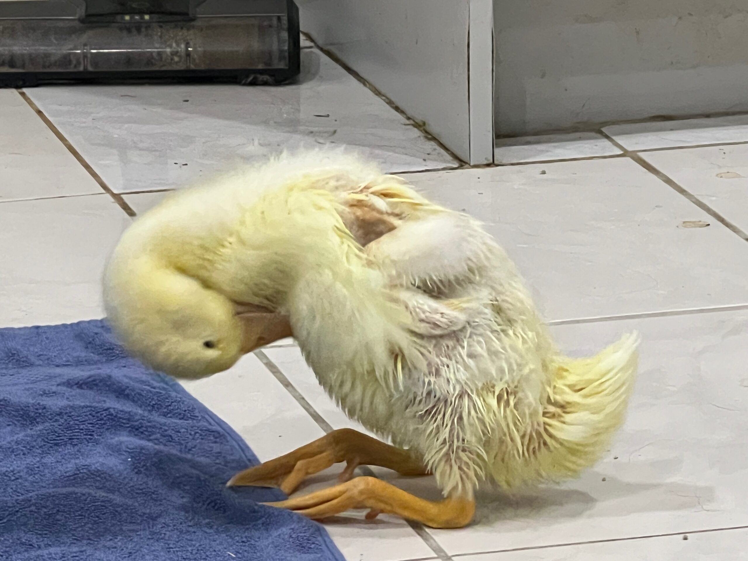
At Bird Vet Melbourne in Burwood, we recently had 4 ducklings present with significant issues caused by malnutrition. Three out of the four, were unable to walk or swim. Two were even unable to stand. All 4 were a fifth of the size they should have been at the age they came in.
Over the next few weeks, our staff worked closely with the birds. Part of the treatment was correcting the nutritional deficiencies.
We saw (and treated) a range of symptoms including seizures, intention tremors and movement difficulties. All of the birds undertook physiotherapy and laser therapy to help correct these issues.
Medications were used to treat the seizures and other abnormalities detected in a blood panel. (Some raised kidney and liver enzymes were found.)
As the birds grew older, two birds were clearly struggling more. They displayed intention tremors and had exaggerated movements when walking, running and swimming. One bird stood out more than the others. He would fall a lot, and his gait was uncordinated. At the time that the CT scan was performed he had no wing movement at all, would fall regularly and seem to lose most of his movement after a seizure.
The question was why? A CT scan would assist with the diagnosis. We wanted to check for cerebellar hypolasia or to see if there was some other cause.
Peparing for A Bird CT
Ideally a patient needs to stay very still during the scanning process. This means that a general anaesthetic is usually required.
Duck anaesthetics are challenging in part because ducks have an incredible ability to hold their breath. Using any sort of gas will require patience! Getting the position of the patient right and the level of anaesthesia will take a few minutes.
During the Bird CT
The anaesthetic nurse monitors the patient, making sure it doesn't move but remains stable. In general, bird anaesthetics need to be fairly light in order for the safety of the patient and to ensure a faster recovery. In this instance a full CT scan of the bird's body was done without contrast, and then another with contrast. This was all done on site at the Burwood Vet clinic with the team from Insight.
Below: An extract of the CT scan process pre-administration of the contrast.
Below: An extract of the CT scan process after contrast has been given.
The Full CT Scan
The Radiography team monitor the quality of the images on their laptop. The images taken are analysed in their complete form later, which shows the 3D version of the scan. A report is compiled and analysed by a specialist and returned to the clinic/owner.
When the scan is completed, the bird is placed into a heated incubator to recover from the anaesthetic. Administration of fluids, is important at this stage.
CT Scan Results
The CT Scan report came back with the following results:
"Both cerebral hemispheres have a normal volume and are symmetric (orange arrows). The 4th ventricle and recess of the 4th
ventricle are mildly prominent (green arrows), while the remaining ventricular system is within normal limits, without evident signs
of distention. However, the cerebellum seems to be normal in size and shape (pink arrows). The intracranial structures have
normal attenuation, without evident abnormal areas of enhancement. There are no signs of mass effect, with no midline shift, and
without evident signs of herniation.
The bones of the skull are within normal limits without evident signs of fractures.
There are no evident abnormalities in the extracranial structures partially included (part of the nasal cavity, sinuses, structures of
the ears, nasopharyngeal duct and ocular globes and orbits).
Conclusions
1. Very mildly prominent 4th ventricle which could be suggestive of very subtle cerebellar hypoplasia. However, this could be an anatomical variation since the cerebellum has normal shape.
2. Otherwise, study of the head unremarkable."

What Was The Resulting Treatment?
Knowing that the brain changes were mild, the Bird Vet Melbourne avian vets working with Winchester opted for a range of treatments to support Winchester's mobility development. Winchester undertook physiotherapy, and photobiomodulation (laser) therapy. He was also put on medication to support his liver (abnormalities were detected in his blood results).
The results were great. Present day, he's off medication and appears happy and healthy. His gait is sometimes slightly exaggerated but he rarely falls and has regained full wing movement.
